Assistant Professor New York Institute of Technology, United States
Purpose: Anatomy lab is the first time medical students are exposed to the complexities of anatomy, often via cadaveric dissections mixed with radiology. Unfortunately, accessing organs such as the heart, requires the destruction of the intimate connections to other anatomical structures. We sought to create a protocol that accurately captured soft-tissue data from in-situ, embalmed organs using Diffusible Iodine-based Contrast-Enhanced Computed Tomography (diceCT).
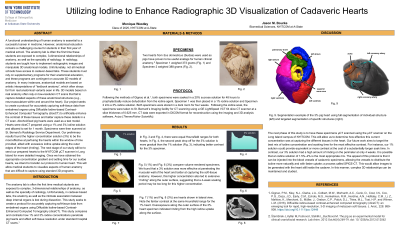
Methods/Materials: We used two fixed pig hearts as proxies for human hearts. Specimens were prophylactically soaked in a 20% sucrose solution for 48 hours to reduce dehydration. Specimen 1 was placed in a 1% iodine solution. Specimen 2 was soaked in a 3% iodine solution and both were stored in the dark for 4 weeks prior to CT scanning. Specimens were scanned at a slice thickness of 0.625 mm. CT data were exported as DICOMs for import and digital reconstruction using the 3D analysis software, Avizo.
Results: The reconstruction window for both hearts generated two high-value peaks. The two specimens differed in the decay rate of the second Hounsfield peak with Specimen 2 showing a more gradual decline indicating a greater contrast and better separation of soft tissues in Specimen 2. However, overexposure along the outer surface of Specimen 2 indicated that oversaturation (rinding) started to occur. 3D models of these contrast-enhanced hearts were able to capture some of the nuances of each heart.
Conclusions: Specimen 2 (3% Iodine solution) had more effective penetration of the muscular walls. However, extensive “rinding” observed along the heart perimeter indicated oversaturation in this area. Based on our results, preserved hearts under a 3% diceCT treatment should not be soaked longer than 4 weeks. Unlike the artistic interpretations commonly available to medical students for showcasing 3D heart anatomy, our approach captured the real, variable anatomy of these hearts at a relatively cheap cost. The next stage of our study is to directly infuse in situ cadaveric hearts using the coronal arterial network. This should provide the same (if not greater) level of contrast without disturbing the natural position of the heart inside the body cavity.